
Cryo-TEM Characterization for Emerging and Specialized Nanoparticle Platforms
Per-Particle Imaging for Formulations That Don't Fit Standard Playbooks
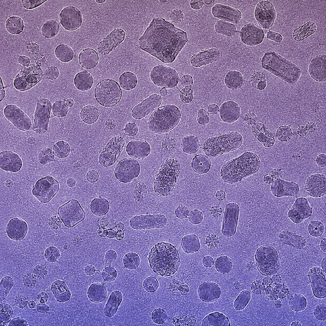
From discovery to the clinic, method validation and regulatory filings, cryo-TEM and negative stain TEM per-particle imaging complements your nanoparticle characterization toolkit at any stage of the pipeline. Cryo-TEM imaging uncovers critical quality attributes (CQAs) that other techniques may overlook, providing a deeper understanding of your nanoparticles, all with only ~50μl of sample.
Platforms We Support
Extracellular Vesicles & Exosomes
Cryo-TEM can provide detailed insights into exosome and extracellular vesicle formulations in their near-native state. Direct visualization of exosomes and EVs allows insights into complex formulations to inform lot comparisons, batch-to-batch scale up, and stability studies.
Cryo-TEM or NS TEM reveals multiple characteristics about exosomes and EVs:
- Size distribution
- Structure
- Lamellarity
- Cargo encapsulation
- Surface decoration
Quantitative and qualitative data, along with method validated studies, provides a deeper understanding of EV and exosome formulations.
Bacteriophages
Cryo-TEM imaging and analysis provides per-particle characterization of bacteriophage formulations, including morphology, size distribution, structural integrity, and aggregation assessment. Validated methods and data can inform batch-to-batch variability, scale-up consistency, and process development changes.
Polymeric Nanoparticles
Nanoparticle size, shape, lamellarity, and morphology affect drug incorporation, stability, and release, which in turn affect cell toxicity, targeting, and therapeutic efficacy. Cryo-TEM imaging provides quantitative and qualitative insights into polymeric nanoparticle formulations in a single study.
Inorganic Nanoparticles
Cryo-TEM imaging and automated image analysis tools can assess iron nanoparticle CQAs including particle size distribution, dense core size, morphology, and aggregation. Iron nanoparticle formulations can have sizing analysis performed on both the particles and dense cores.
Protein-Based Nanoparticles
Self-assembling protein nanoparticles, such as ferritin cages and encapsulins, can display numerous proteins, peptides, or antigens on their surface depending on the application. Characterizing the surface protein spatial distribution using cryo-TEM classifies particles as densely, sparsely, or minimally decorated, supporting nanoparticle platform optimization across vaccine, drug delivery, and diagnostic applications. Cryo-electron tomography (cryo-ET) provides 3D volumes to better distinguish surface protein distribution and internal particle features.
What Cryo-TEM Reveals Across All Platforms
A single cryo-TEM imaging study can simultaneously provide information on:
- Particle size distribution
- Drug and payload encapsulation and distribution
- Morphology, including shape, circularity, lamellarity, and blebbing
- Aggregation
- Interactions with and effects of adjuvant
- Sample integrity and impurities
- Surface decoration and protein distribution
- 3D volumes with cryo-electron tomography