
Cryo-EM Structure of CYP3A4: Unliganded and Substrate-Bound
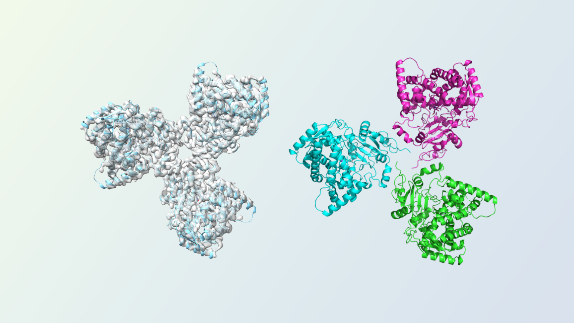
This TrueCourse white paper presents a gene-to-structure case study of the CYP3A4 protein in its unliganded form and in complex with hydroxychloroquine, or HCQ. The work combines recombinant protein production, analytical characterization, cryo-EM data collection, and structure interpretation to examine a 57 kDa drug-metabolizing enzyme.
Complete the form to download the white paper and review the study workflow, preferred-orientation strategy, unliganded reconstruction, HCQ-bound reconstruction, and structural interpretation.
Download the CYP3A4 Cryo-EM White Paper
Submit the form to receive “Cryo-EM Structure of CYP3A4 Unliganded and In Complex with a Substrate.”
CYP3A4 Protein: Structure and Study Context
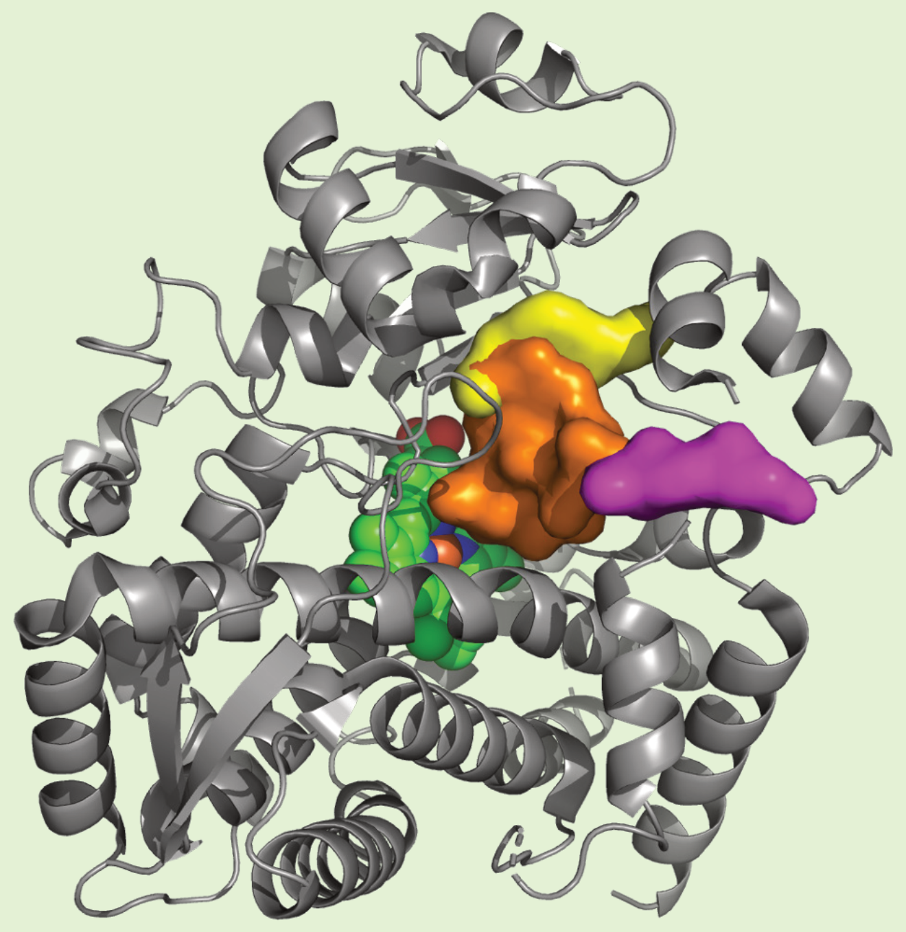
CYP3A4 is a heme-containing cytochrome P450 enzyme with a broad substrate range and a central role in drug metabolism. Structural studies have described a flexible active region, a substrate access channel, and a peripheral binding site. These features help frame questions about substrate recognition, inhibition, conformational behavior, and higher-order assembly. [1,2]
The white-paper study focuses on the soluble domain of CYP3A4. Recombinant protein was expressed in E. coli and purified using Ni-NTA affinity chromatography, ion exchange chromatography, and preparative size-exclusion chromatography. SDS-PAGE, HPLC-SEC, and intact mass spectrometry were used to assess the final preparation.
Why use cryo-EM for a 57 kDa CYP3A4 target?
CYP3A4 has an extensive crystallographic record, yet cryo-EM can provide a complementary view of particle behavior in solution, including flexibility, oligomeric state, and orientation. In this study, negative-stain and cryo-EM class averages supported the presence of oligomeric particles, with a trimer as the most common observed assembly. The functional relevance of the cryo-EM assembly requires further study, although prior membrane-focused work supports the possibility that CYP3A4 oligomerization can influence function. [3]
CYP3A4 Structure by Cryo-EM
The case study reports cryo-EM reconstructions for unliganded CYP3A4 and for CYP3A4 prepared with HCQ. The two datasets were collected on a TFS Titan Krios transmission electron microscope operated at 300 kV. The system included a Gatan Quantum 967 LS imaging filter and a Gatan K3 Direct Detection Camera, with Leginon used for data collection.
Unliganded and hydroxychloroquine-bound CYP3A4
The unliganded dataset was processed in CryoSPARC v4.6.2. Model building began from PDB entry 1W0E and used ChimeraX for map fitting, followed by iterative work in PHENIX v1.19.2 and COOT v0.9.6 EL. PISA was used to evaluate the reported interfaces within the trimer.
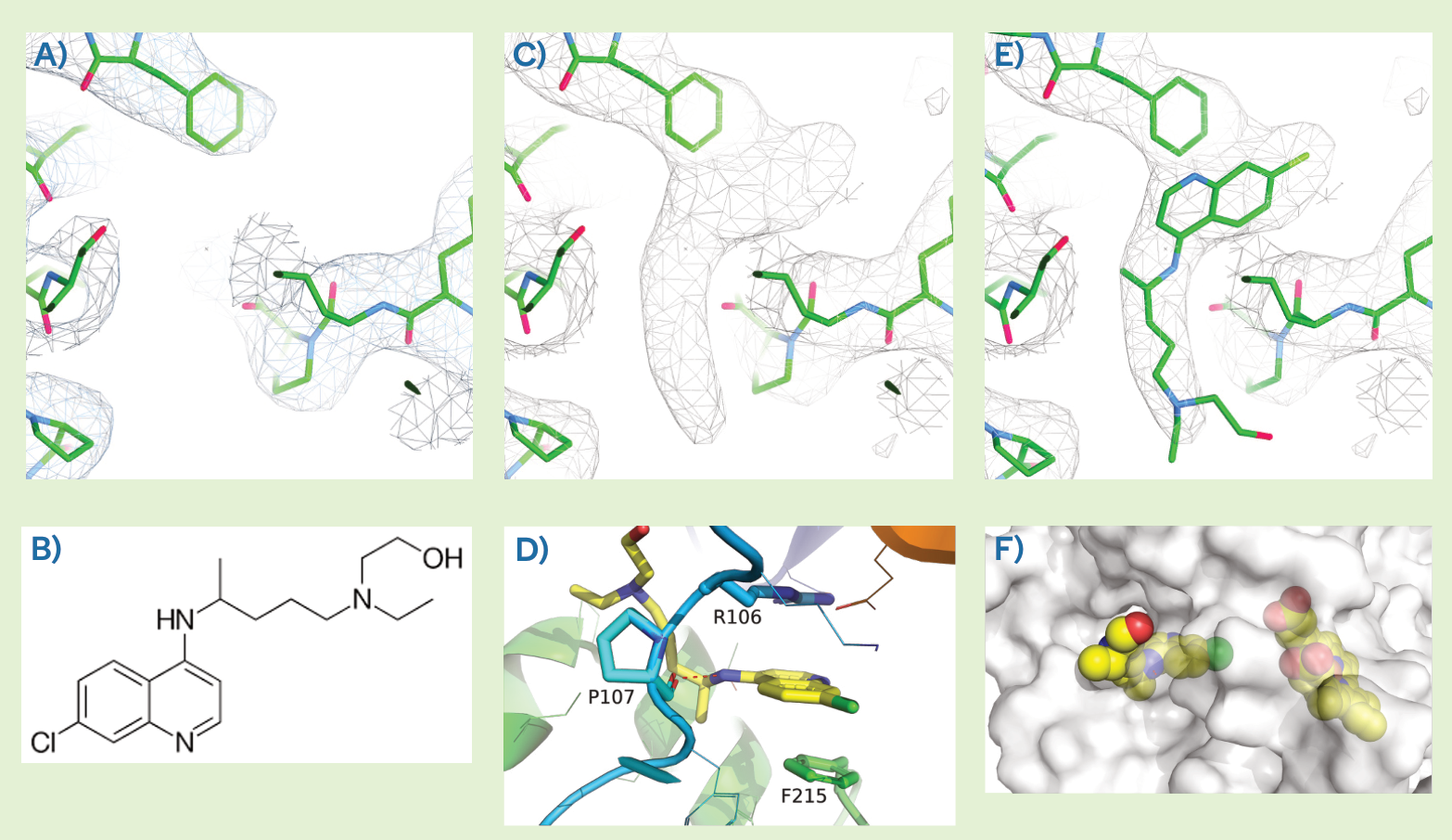
For the HCQ-bound reconstruction, additional density was observed in the substrate access channel. The modeled ligand places the quinoline ring between Phe215 and Arg106, with a hydrogen bond between the quinoline nitrogen and the main-chain oxygen of Pro107. The observed pose was interpreted as nonproductive and discussed in relation to published metabolism and inhibition data. [4,5]
Addressing Preferred Orientation in the Cryo-EM Workflow
Initial vitrification conditions produced strong preferred orientation. The reported structures used tilted data collection, with a 30-degree tilt for the unliganded dataset and a 20-degree tilt for the HCQ-bound dataset. While tilt can improve angular coverage, the thicker ice, beam-induced movement, focus gradients, and particle overlap can complicate collection and processing. [6,7]
The study also screened protein concentration, grid type, and additives. Short collections of approximately 1,200 images on a Glacios microscope were used to assess suitable conditions. LMNG and fluorinated fos-choline-8 were reported as the two most effective tested conditions for reducing orientation bias in this sample. These results are specific to the CYP3A4 preparation and should be evaluated rather than assumed for other proteins.
How a cryo-EM CRO can support a gene-to-structure study
A coordinated workflow can connect expression strategy, purification, analytical characterization, grid screening, data collection, image processing, and model interpretation. For small or orientation-prone targets, early sample feedback and condition screening can help determine whether a high-resolution campaign is scientifically justified.
What You Will Find in the White Paper
- Background on CYP3A4 protein structure and its relevance to drug metabolism.
- The recombinant expression, purification, and analytical characterization workflow.
- Negative-stain and cryo-EM observations of CYP3A4 oligomeric states.
- The preferred-orientation challenge and the collection and screening strategies evaluated.
- The unliganded CYP3A4 reconstruction and structural interpretation.
- The HCQ-bound reconstruction, ligand density, and proposed binding pose.
- Data-collection and refinement statistics for both structures.
White Paper Authors
Claudio Catalano, Lindsey Gottler, Vincent Shaw, Jason Haineault, Kevin Cannon, Ashlyn Farwell, Kyle Lucier, Reid Luksic, Arnold Muccini, and Skerdi Senko.
Frequently Asked Questions
What is the CYP3A4 protein?
CYP3A4 is a human cytochrome P450 enzyme involved in the metabolism of many structurally diverse compounds. It contains a heme cofactor and has a flexible binding environment that can accommodate substrates, inhibitors, and modulators. [1,2]
What is the structure of CYP3A4?
CYP3A4 adopts the characteristic cytochrome P450 fold and contains a heme-centered active region connected to a substrate access channel. Crystallographic studies have also identified a peripheral binding site. The white-paper case study adds cryo-EM observations of a symmetric trimer in the analyzed soluble-domain preparation. [1,2,3]
What does CYP3A4 stand for?
CYP3A4 refers to cytochrome P450 family 3 subfamily A member 4. It is commonly described as CYP3A4 in structural biology, drug metabolism, and pharmacology literature.
Can cryo-EM be used to study a 57 kDa protein?
The reported CYP3A4 case study shows that cryo-EM can be applied to a 57 kDa target when sample behavior, oligomerization, grid conditions, orientation, collection strategy, and processing are favorable. The result should be treated as a case-specific demonstration rather than a universal mass threshold.
Where did hydroxychloroquine bind in the CYP3A4 structure?
In the reported reconstruction, the modeled HCQ density is located in the substrate access channel. The pose places the quinoline ring between Phe215 and Arg106 and was interpreted as a nonproductive orientation.
Why was tilted data collection used for CYP3A4?
Tilted collection was used to address strong preferred orientation and improve angular coverage. The unliganded and HCQ-bound datasets used different reported tilt angles. Tilt can introduce additional collection and processing challenges, so the strategy should be selected in the context of the sample and grid behavior. [6,7]
How do I download the CYP3A4 white paper?
Complete and submit the form on this page. The approved PDF will be delivered through the TrueCourse confirmation or email workflow.
Download the CYP3A4 Cryo-EM Case Study
Access the complete white paper for the protein-production workflow, cryo-EM strategy, structure figures, and reported collection and refinement details.
References
Primary and peer-reviewed sources supporting the scientific context:
1. Williams PA, et al. Crystal structures of human cytochrome P450 3A4 bound to metyrapone and progesterone. Science. 2004;305(5684):683-686. Source
2. Sevrioukova IF, Poulos TL. Understanding the mechanism of cytochrome P450 3A4: recent advances and remaining problems. Dalton Transactions. 2013;42:3116-3126. Source
3. Davydov DR, Davydova NY, Sineva EV, Halpert JR. Interactions among cytochromes P450 in microsomal membranes: oligomerization of cytochromes P450 3A4, 3A5, and 2E1 and its functional consequences. Journal of Biological Chemistry. 2015;290(6):3850-3864. Source
4. Paludetto MN, et al. Hydroxychloroquine is metabolized by cytochrome P450 2D6, 3A4, and 2C8, and inhibits cytochrome P450 2D6, while its metabolites also inhibit cytochrome P450 3A in vitro. Drug Metabolism and Disposition. 2023;51(3):293-305. Source
5. Sevrioukova IF. Crystal structure of CYP3A4 complexed with Fluorol identifies the substrate access channel as a high-affinity ligand binding site. International Journal of Molecular Sciences. 2022;23(20):12591. Source
6. Tan YZ, et al. Addressing preferred specimen orientation in single-particle cryo-EM through tilting. Nature Methods. 2017;14:793-796. Source
7. Aiyer S, et al. Overcoming resolution attenuation during tilted cryo-EM data collection. Nature Communications. 2024;15:389. Source