Cryo-EM Epitope Mapping Guide for Antibody-Antigen Complexes
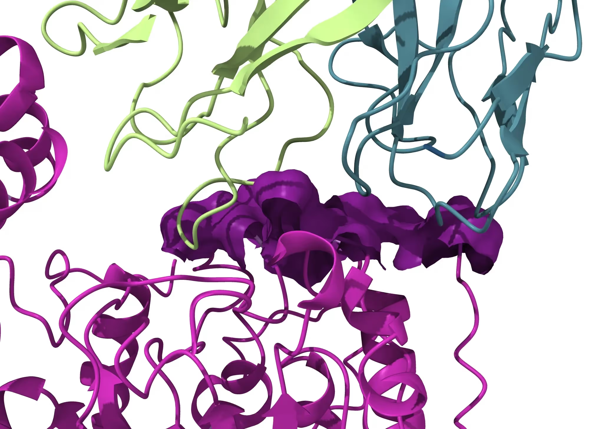
Epitope mapping identifies and characterizes the region of an antigen recognized by an antibody. At sufficient structural detail, it can also describe the antibody paratope and the residue-level interface between the two binding partners. These data can inform antibody optimization, specificity and cross-reactivity assessment, diagnostic development, vaccine research, and the documentation used to support patent strategy [1-4].
No single method answers every epitope question. Cryogenic electron microscopy, X-ray crystallography, hydrogen-deuterium exchange mass spectrometry, and deep mutational scanning provide different kinds of evidence. Method selection should follow the scientific question, sample behavior, desired structural detail, material constraints, and project timeline.
Epitope Mapping Services
An epitope mapping service should begin by defining the decision the study must support. A project seeking residue-level interpretation of an antibody-antigen complex has different requirements from a project seeking a broad interaction region, a competition group, or a screen across many variants. The service plan should connect the method, sample requirements, controls, intermediate outputs, and reporting format to that decision.
- Confirm whether the desired output is a binding region, competition bin, three-dimensional interface, or residue-level model.
- Review the size, ordered mass, flexibility, affinity, homogeneity, buffer, available concentration, and available sample volume.
- Specify how preferred orientation, dissociation, aggregation, or inadequate vitrification will be assessed and addressed.
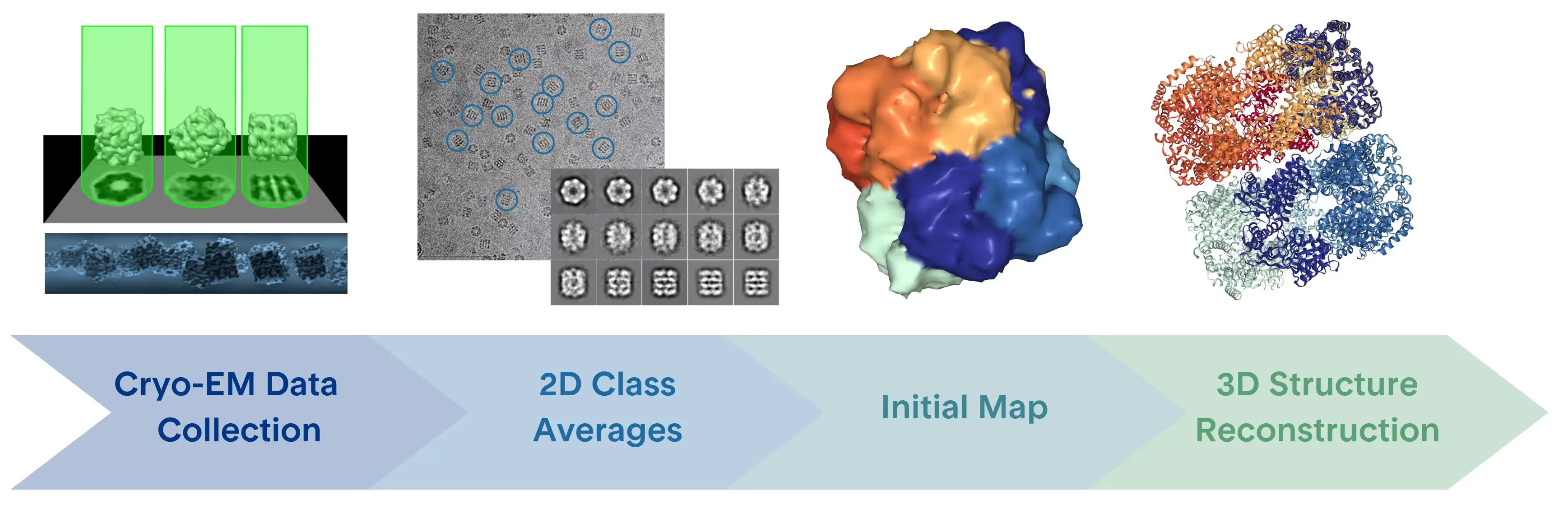
- Define interim checkpoints such as screening micrographs, 2D class averages, initial maps, and a decision to continue or optimize.
- Request explicit reporting of sample conditions, instrument configuration, processing software, map quality, model confidence, and study limitations.
What Is Epitope Mapping?
The epitope is the region of an antigen recognized by an antibody. The paratope is the complementary binding surface on the antibody. Epitope mapping can range from localizing a general binding region to resolving the three-dimensional arrangement and specific amino acid interactions at the interface.
Epitope mapping can inform immune-response specificity, cross-reactivity, and immunogenicity, as well as antibody and vaccine development, diagnostic design, and patent filings. The depth of the conclusion depends on the technique and the quality of the resulting evidence [1-4].
Antibody Epitope Mapping and Paratope Mapping
Antibody epitope mapping focuses on the antigen surface recognized by the antibody. Paratope mapping focuses on the antibody residues that engage the antigen. A three-dimensional structure of the complex can support both views when the map and model provide sufficient local detail. Lower-resolution or indirect methods may localize an interaction region without defining every residue-residue contact.
Conformational Epitope Mapping
Antibody-antigen recognition often involves discontinuous, or conformational, epitopes. These epitopes are formed by residues that are close in the folded three-dimensional structure even when they are separated in the primary sequence. Their recognition can therefore depend on the antigen retaining the relevant conformation.
Cryo-EM and X-ray crystallography can visualize a three-dimensional binding interface. HDX-MS and deep mutational scanning can provide complementary evidence about protected regions, conformational change, or the functional effect of mutations. The methods should be interpreted according to what each measurement directly observes [1, 6].
Epitope Mapping Methods and Techniques
The methods provide different structural or functional evidence and should be interpreted according to the signal each technique directly measures [1, 2, 6].
High-Resolution 3D Epitope Mapping with Cryo-EM
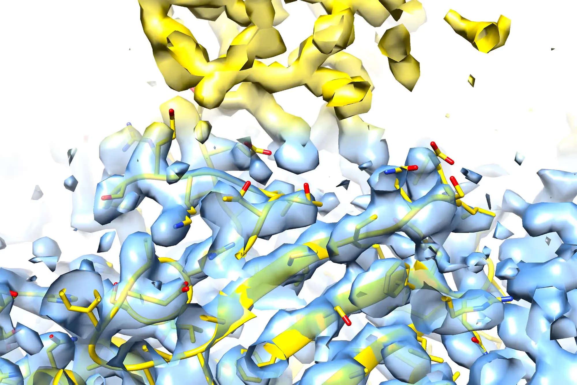
Approximately 3.5 angstroms can serve as a practical planning target for residue-based interpretation at the epitope-paratope interface. For a well-behaved sample, the workflow may target 3.5 angstroms or better with chain tracing and assignment of 75% of side chains. These are project benchmarks rather than universal outcomes.
At sufficiently resolved interfaces, a cryo-EM model may help distinguish antibodies with similar epitopes, identify conformationally specific antibodies, inform rational in silico design, and show surface features such as post-translational modifications near the interface. Interpretation should remain tied to local map quality and model confidence.
Practical ordered-mass guidance
Practical mass guidance depends on complex behavior and the amount of ordered density available for alignment. An antigen contributing more than 50 kDa of ordered mass, combined with an approximately 52 kDa Fab, can create a local ordered mass of about 100 kDa around the interface. Antigens much smaller than 50 kDa may still be mapped in some circumstances. A routine antibody-antigen or Fab candidate may have approximately 80 to 100 kDa of ordered mass, while orientation assignment can become difficult below 80 kDa. These values should be treated as planning heuristics rather than universal cutoffs.
Fab fragments, full-length antibodies, and multiplexed complexes
Cryo-EM epitope mapping can use both Fab-antigen complexes and full-length antibodies. Fabs can reduce sample complexity and focus the ordered mass around the interface. Full-length antibodies may add mass and sometimes reduce preferred orientation, but their flexibility, aggregation, or extended chains can complicate vitrification and reconstruction. Fab fragments generated by digestion of a monoclonal antibody can avoid recombinant Fab production.
When several non-competing antibodies bind unique sites, a multiplexed complex may increase particle mass and reduce preferred orientation. Individual epitopes can then be interpreted during processing and three-dimensional reconstruction, provided the sample and data support separation of the interfaces.
Affinity, buffer, concentration, and preferred orientation
- Affinity: higher vitrification concentrations can accommodate weaker pairs than some negative-stain workflows, while high-affinity, homogeneous systems generally reduce workflow complexity.
- Buffer: glycerol and sucrose can increase background signal, and DMSO can interfere with vitrification. Effects depend on composition and concentration.
- Sample amount and concentration: 50 to 100 microliters at 0.5 to 5 mg/mL is practical guidance for imaging in this workflow. Samples are used at micromolar concentration.
- Dissociation: a complex with a dissociation constant worse than micromolar may tend to dissociate at the concentrations used in this workflow.
- Preferred orientation: detergents, changes in grid substrate, different vitrification methods, and tilted data collection are possible responses. Preferred orientation was observed in approximately half of the Fab cases evaluated by the team.
How Do I Do Epitope Mapping with Cryo-EM?
A cryo-EM epitope-mapping workflow for a well-behaved antibody-antigen sample can follow the sequence below. Each step is a decision point, and samples that show instability, preferred orientation, inadequate ice, or limited structural signal may require additional optimization.
- Optimize and screen the sample. Apply an aliquot to a grid and vitrify it in liquid ethane, aiming for thin, uniform ice with particles in multiple orientations.
- Screen grids for suitability. TrueCourse can screen grids on one of four Thermo Fisher Scientific Glacios microscopes and adjust detergents, grid types, or vitrification methods when needed.
- Collect an initial data set. Overnight collection on a Glacios microscope can proceed with cryoSPARC Live processing in parallel.
- Review 2D classifications. The next-morning classes help assess secondary-structure visibility, orientation diversity, and whether the sample should move forward.
- Generate optimized 2D class averages and an initial map. Further processing over the next few days can expose preferred orientation or other limitations more clearly.
- Collect and process the larger data set. If the initial map supports continuation, additional microscope time and refinement are used to pursue a high-resolution three-dimensional reconstruction.
Workflow timeline and instrumentation
For a well-behaved antibody-antigen pair, a rapid first-pass workflow may yield a 3 to 3.5 angstrom epitope map within 24 to 48 hours from the start of an experiment. Instrument configurations include a Thermo Fisher Scientific Glacios with a Falcon 4 detector and a Thermo Fisher Scientific Krios with a K3 detector, with cryoSPARC Live used for processing. A fully refined three-dimensional reconstruction may be completed within two weeks of sample receipt for a well-behaved sample. These timelines are workflow examples and should not be generalized to every sample or project.
Epitope Mapping for Drug Discovery
Structural epitope information can support several drug-discovery decisions. It can help compare antibody specificity, evaluate cross-reactivity, identify conformationally specific binding, guide sequence and structure-based optimization, and provide evidence for the novelty and specificity described in patent filings [3, 4].
The appropriate level of detail depends on the decision. A broad interaction region may be sufficient for candidate triage, while residue-level interpretation may be needed to compare closely related binding modes or document a distinctive interface. Method choice should therefore be made before the study is scoped, with explicit consideration of the sample and the evidence required.
.avif)
Frequently Asked Questions
What is epitope mapping?
Epitope mapping identifies and characterizes the region of an antigen recognized by an antibody. Depending on the method, the output can range from a broad binding region to a three-dimensional, residue-level interface.
How do I do epitope mapping?
Start with the decision the result must support, then select a method that provides the required structural or functional detail. For cryo-EM, the workflow typically includes sample assessment, grid optimization, vitrification, screening, 2D classification, an initial map, expanded data collection, and three-dimensional reconstruction.
How does epitope mapping guide antibody optimization and sequence refinement?
A sufficiently detailed interface can show which antigen and antibody residues contribute to binding, reveal nearby structural features, and help compare candidate binding modes. Those observations can guide hypotheses for sequence changes, which should then be tested experimentally.
Which approaches are best for studying antibody-antigen interactions at high resolution using electron microscopy?
Cryo-EM single-particle analysis can support high-resolution three-dimensional epitope mapping. Suitability depends on ordered mass, particle behavior, orientation distribution, vitrification, data quality, and the local detail needed at the interface.
How do I choose an epitope mapping service provider?
Choose a provider that can connect the method to the scientific decision, assess sample suitability before full data collection, explain intermediate go or no-go criteria, report the instrument and processing workflow, and qualify resolution and model claims at the local interface.
What is the difference between epitope mapping and epitope binning?
Epitope mapping aims to localize or structurally define an antibody's binding site. Epitope binning groups antibodies according to competition or overlap patterns, often without identifying the exact residues. Binning can help organize a panel before higher-resolution mapping [7].
Can cryo-EM map a conformational epitope?
Cryo-EM can visualize a folded antibody-antigen complex and may resolve a conformational epitope when the particles, data, map, and local interface detail support the interpretation. Flexible loops or poorly ordered regions can remain difficult to model.
Discuss a Cryo-EM Epitope Mapping Study
TrueCourse can help evaluate whether cryo-EM, X-ray crystallography, HDX-MS, deep mutational scanning, or a staged combination is appropriate for an antibody-antigen question. Planning should account for sample behavior, ordered mass, flexibility, affinity, buffer, available material, desired structural detail, and the decision the study must support.
A related webinar presented by Dr. Giovanna Scapin, CSO, discusses epitope mapping with cryo-EM and how the method compares with other approaches.
Peer-reviewed references cited in the draft
[1] Francino-Urdaniz, I. M., & Whitehead, T. A. (2021). An overview of methods for the structural and functional mapping of epitopes recognized by anti-SARS-CoV-2 antibodies. RSC Chemical Biology, 2(6), 1580-1589. https://doi.org/10.1039/D1CB00169H
[2] Malito, E., Carfi, A., & Bottomley, M. J. (2015). Protein Crystallography in Vaccine Research and Development. International Journal of Molecular Sciences, 16(6), 13106-13140. https://doi.org/10.3390/ijms160613106
[3] Gershoni, J. M., Roitburd-Berman, A., Siman-Tov, D. D., Tarnovitski Freund, N., & Weiss, Y. (2007). Epitope mapping: the first step in developing epitope-based vaccines. BioDrugs, 21(3), 145-156. https://doi.org/10.2165/00063030-200721030-00002
[4] Deng, X., Storz, U., & Doranz, B. J. (2018). Enhancing antibody patent protection using epitope mapping information. mAbs, 10(2), 204-209. https://doi.org/10.1080/19420862.2017.1402998
[5] Nikitin, P. A., DiMuzio, J. M., Dowling, J. P., et al. (2022). IMM-BCP-01, a patient-derived anti-SARS-CoV-2 antibody cocktail, is active across variants of concern including Omicron BA.1 and BA.2. Science Immunology, 7(75), eabl9943. https://doi.org/10.1126/sciimmunol.abl9943
[6] Dang, X., Guelen, L., Lutje Hulsik, D., et al. (2023). Epitope mapping of monoclonal antibodies: a comprehensive comparison of different technologies. mAbs, 15(1), 2285285. https://doi.org/10.1080/19420862.2023.2285285
[7] Brooks, B. D., Closmore, A., Yang, J., Holland, M., Cairns, T., Cohen, G. H., & Bailey-Kellogg, C. (2020). Characterizing Epitope Binding Regions of Entire Antibody Panels by Combining Experimental and Computational Analysis of Antibody: Antigen Binding Competition. Molecules, 25(16), 3659. https://doi.org/10.3390/molecules25163659
.png)
.png)
.png)