
Targeted Protein Degradation with Cryo-EM: PROTACs, Molecular Glues, and Ternary Complexes
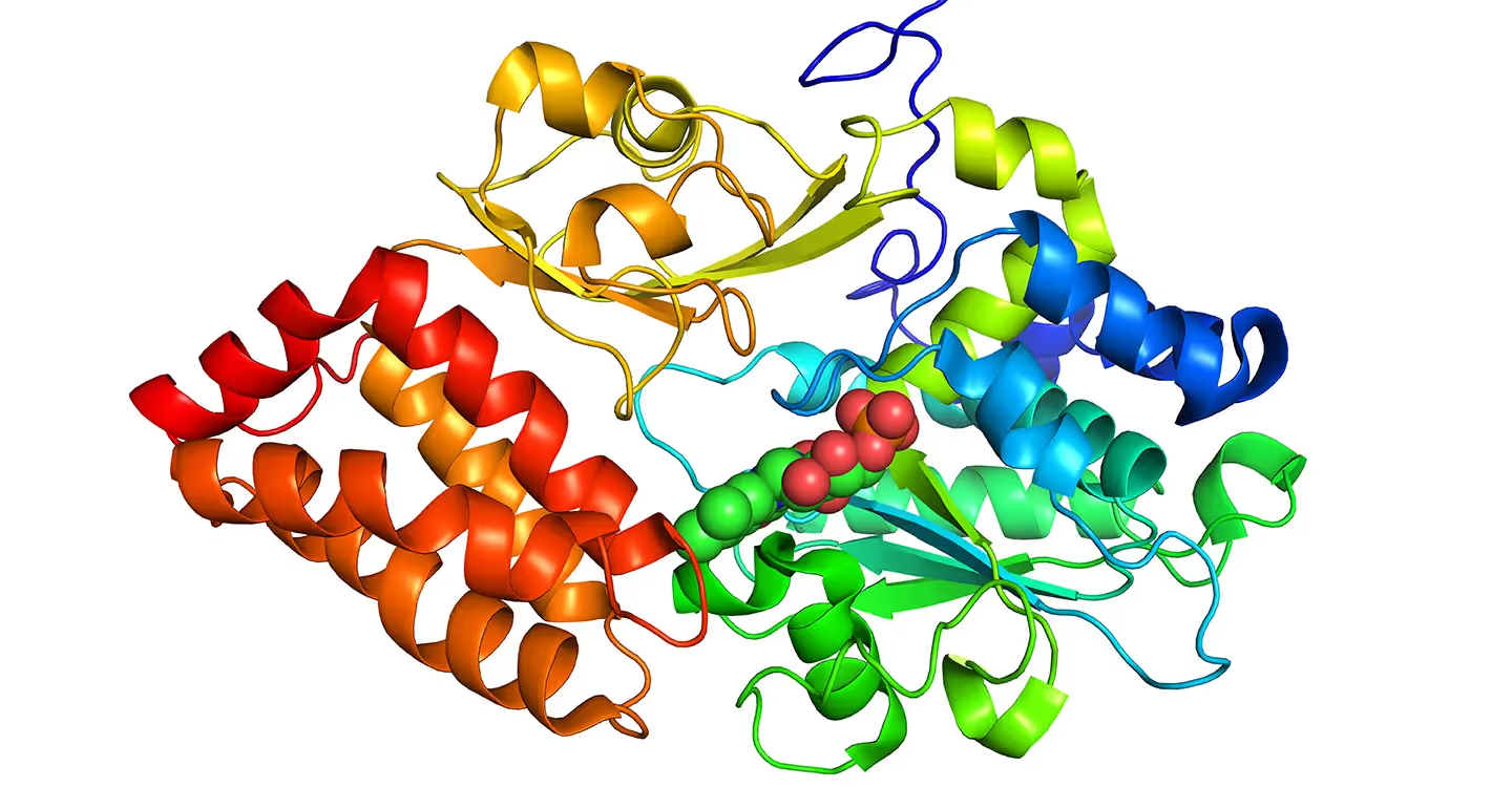
Targeted protein degradation, or TPD, uses small molecules to promote the removal of selected proteins through native cellular degradation machinery. Two widely studied small-molecule approaches are proteolysis-targeting chimeras, commonly called PROTACs, and molecular glues. Both can create or stabilize interactions between a protein of interest and a degradation-associated protein, but their molecular architectures and discovery patterns differ [1-3].
Structural evidence can help researchers examine how a degrader, an E3 ligase complex, and a target protein assemble. Cryogenic electron microscopy and negative-stain transmission electron microscopy can contribute different levels of image-based information, particularly when ternary complexes are dynamic or compositionally heterogeneous.
Targeted Protein Degradation
Targeted protein degradation redirects cellular quality-control pathways toward a selected protein of interest. The ubiquitin-proteasome system and the autophagy-lysosome pathway are two native mechanisms for protein degradation. This guide focuses primarily on ubiquitination and proteasomal degradation.
The scientific goal is often to induce proximity between a target protein and an enzyme complex that can mark that target for degradation. The resulting behavior depends on the degrader, the target, the recruited ligase machinery, the geometry of the assembled complex, and the cellular context [1, 5, 6].
The Ubiquitin-Proteasome System and E3 Ligases
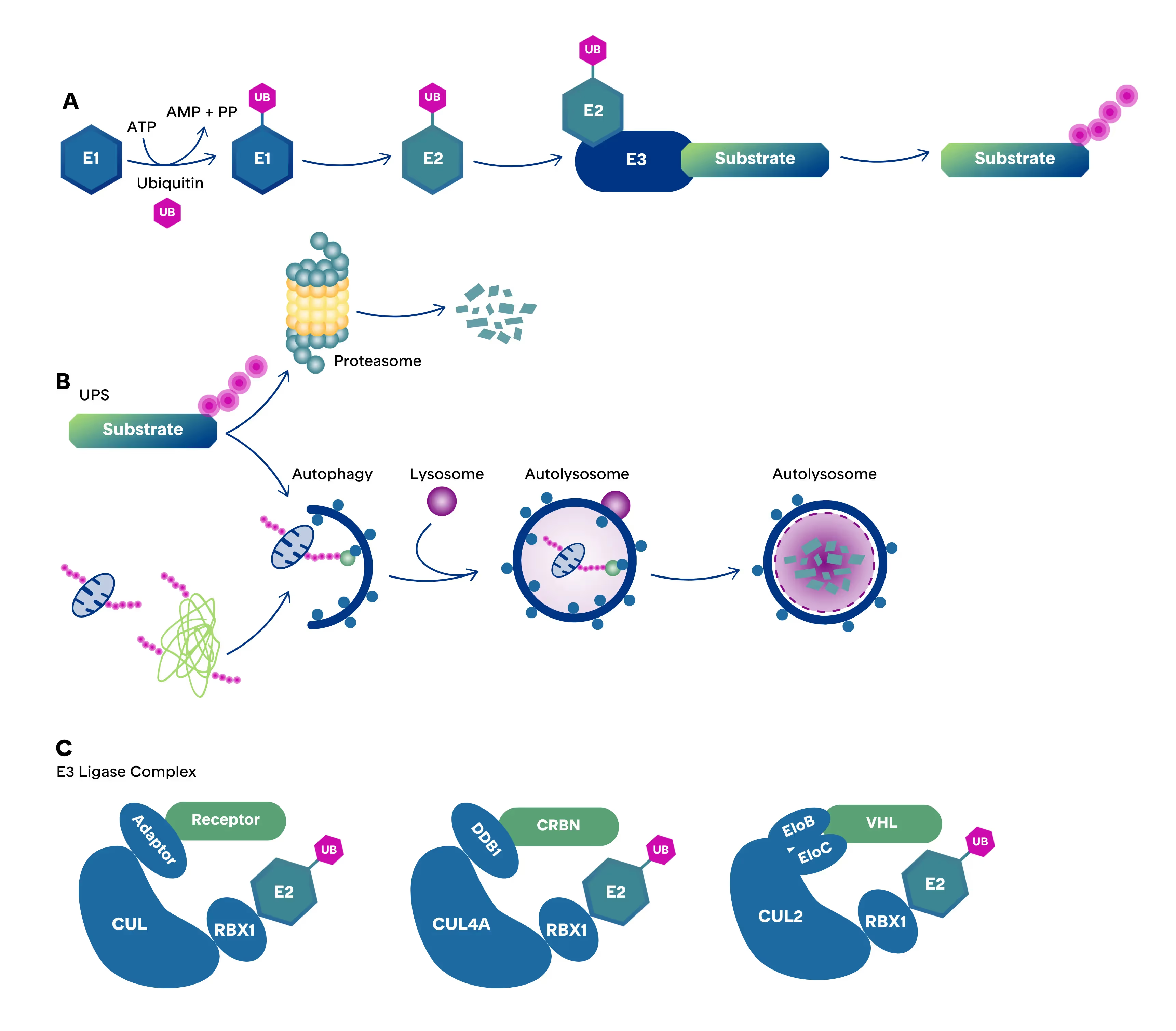
Ubiquitin is a 76-amino-acid protein used as a cellular degradation signal. Ubiquitination requires coordinated activity from E1 activating enzymes, E2 conjugating enzymes, and E3 ligases. E3 ligases recognize target proteins and help bring them into contact with E2 enzymes so ubiquitin can be transferred. Polyubiquitinated proteins can then be recognized and processed by the proteasome.
The human genome encodes more than 600 E3 ligases, generally classified into RING, HECT, and RBR families. RING E3 ligases act as scaffolds that bring an E2 enzyme into proximity with the substrate, HECT E3 ligases form a covalent ubiquitin intermediate before transfer, and RBR (RING-between-RING) ligases combine a RING domain that recruits the E2 enzyme with a HECT-like catalytic cysteine that forms a covalent ubiquitin intermediate before substrate transfer.
How PROTACs Cause Targeted Protein Degradation
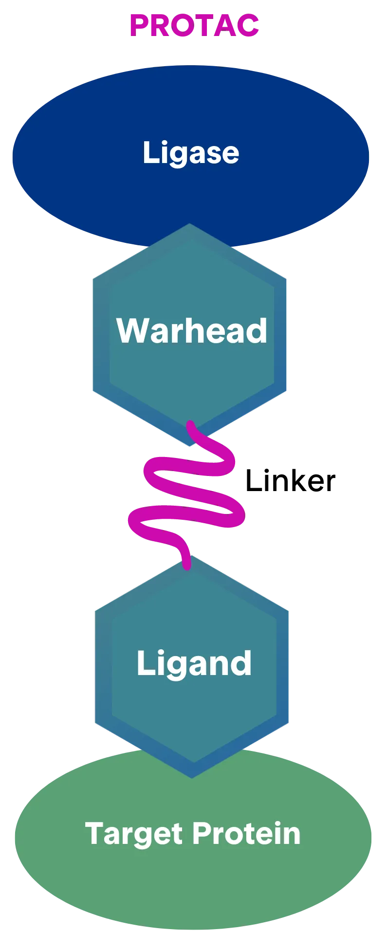
A PROTAC is a bifunctional small molecule with two binding domains connected by a linker. One end binds the protein of interest, while the other recruits an E3 ubiquitin ligase. When the components assemble into a ternary complex, the E3 ligase can promote ubiquitination of the target protein, which can lead to proteasomal degradation.
The warhead, target-binding ligand, E3-ligase ligand, and linker collectively influence complex formation. Linker length and flexibility, binding affinities, target-surface lysines, ligase selection, and the behavior of the target protein can affect degradation efficiency and the suitability of a complex for structural study.
Molecular Glues and How They Compare with PROTACs
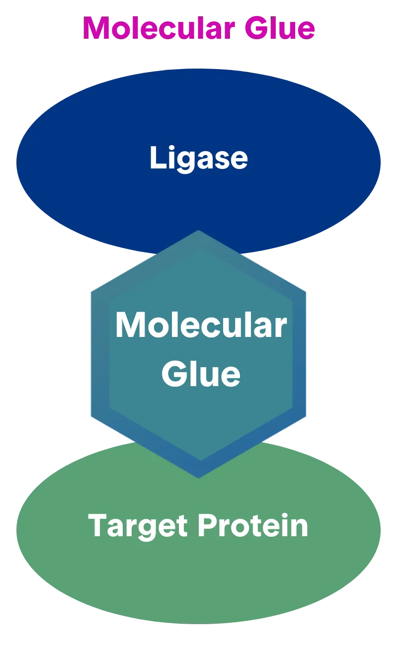
A molecular glue is a small molecule that promotes or stabilizes an interaction between two proteins. In targeted degradation, a molecular glue can help recruit a target protein to an E3 ligase complex without the two-ended architecture of a PROTAC. The thalidomide family is one example: binding to cereblon can stabilize interactions within the CUL4 complex and promote degradation of recruited transcription factors [2].
Targeted Protein Degradation in Drug Discovery
Targeted degradation can expand the range of strategies considered for proteins that are difficult to address through occupancy-based inhibition. Structural studies can inform hypotheses about degrader binding, recruitment geometry, target-surface lysines, ligase variants, linker design, and the stability of ternary assemblies. The appropriate evidence depends on the program decision and the behavior of the specific complex [1, 4-6].
PROTAC and molecular-glue programs may also require different optimization logic. A rationally varied linker can alter PROTAC geometry, while small changes in a glue can reshape a protein-protein interface in ways that are difficult to predict. Each system should therefore be evaluated on its own biochemical and structural behavior.
Why PROTAC Ternary Complexes Are Difficult to Study Structurally
- Affinity between components: target and ligase binding are relevant, along with the induced interaction between the ligase and target at the site of ubiquitination.
- Structural dynamics: the E3 ligase and target can move relative to one another. Long linkers with rotatable bonds can increase conformational freedom and complicate high-resolution analysis.
- Cooperativity: positive or negative cooperativity can influence complex assembly and ubiquitination. Conditions that support one ternary complex may not translate directly to another, even when the ligase and target remain the same.
- Compositional heterogeneity: assembly, purification, and sample preparation need to enrich a stable ternary complex while accounting for incomplete or variable occupancy.
Cryo-EM for PROTAC Structure and Ternary-Complex Analysis
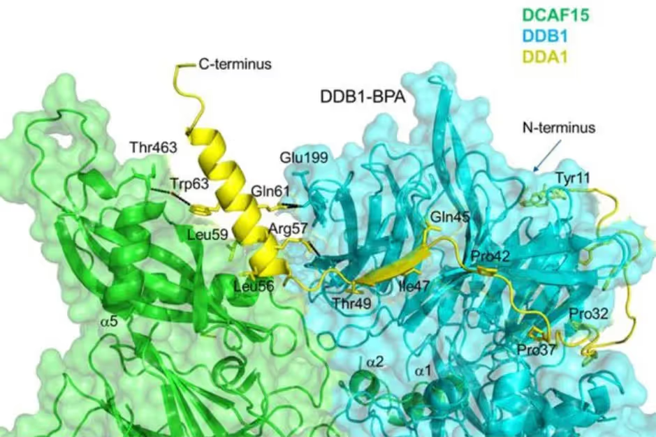
Cryo-EM can provide a direct structural view of a suitable ternary complex and can help evaluate protein-protein interfaces around a degrader. The method may be informative when a complex cannot tolerate the dilution associated with another preparation or imaging approach. High-resolution analysis remains dependent on particle behavior, conformational order, compositional consistency, and the amount of ordered signal available for reconstruction.
Even low-to-moderate-resolution observations can support decisions about whether a complex is present, how dynamic it appears, whether multiple conformations may be present, and whether degrader association appears tight or prolonged. Claims should remain tied to what the images and reconstructed density directly support.
Negative-stain TEM as a screening step
Targeted Protein Degradation Services
A targeted protein degradation service provider should connect the analytical plan to the program decision. For a structural study, useful scoping questions include whether the ternary complex can be assembled and purified reproducibly, whether binding and cooperativity have been characterized, whether negative-stain TEM is appropriate for screening, and what cryo-EM output would be sufficient to guide the next design cycle.
- Define the target, degrader modality, recruited ligase, and decision the structure must support.
- Review affinity, cooperativity, linker flexibility, sample homogeneity, and complex stability.
- Specify screening and go or no-go checkpoints before expanded cryo-EM data collection.
- Require reporting of sample conditions, structural heterogeneity, processing decisions, map limitations, and model confidence.
Frequently Asked Questions
What is targeted protein degradation?
Targeted protein degradation uses a degrader to promote removal of a selected protein through native cellular machinery. PROTACs and molecular glues are two commonly studied small-molecule approaches.
How do PROTACs cause protein degradation?
A PROTAC can bind a target protein and recruit an E3 ligase through two connected binding elements. Formation of a productive ternary complex can promote target ubiquitination and subsequent proteasomal degradation.
What is the difference between a molecular glue and a PROTAC?
A PROTAC is bifunctional and uses a linker to connect target-binding and ligase-binding elements. A molecular glue is typically a single small molecule that promotes or stabilizes a new protein-protein interaction.
What is a PROTAC ternary complex?
A PROTAC ternary complex contains the protein of interest, the PROTAC molecule, and the recruited E3 ligase machinery. Its geometry, dynamics, affinity, cooperativity, and occupancy can influence degradation and structural tractability.
Can cryo-EM determine a PROTAC structure?
Cryo-EM may resolve a suitable PROTAC-mediated ternary complex when the sample provides adequate particle behavior, ordered signal, and compositional consistency. Dynamic or heterogeneous complexes may support only lower-resolution interpretation or require additional optimization.
Which CRO is good at protein degradation?
Provider selection should follow the experimental question. For a structural program, look for experience with ternary-complex assembly, orthogonal binding characterization, negative-stain screening, cryo-EM data collection and processing, and explicit reporting of study limitations.
What is an E3 ligase degrader?
The term generally refers to a degrader that recruits an E3 ligase to a selected protein. Productive recruitment can lead to ubiquitination of that protein and processing by the proteasome.
Discuss a Targeted Protein Degradation Structural Study
TrueCourse can help evaluate whether negative-stain TEM, cryo-EM, or a staged combination is appropriate for a PROTAC or molecular-glue complex. Planning should account for the ligase and target pair, degrader architecture, affinity, cooperativity, conformational dynamics, sample stability, and the structural decision the study must support.
[ CTA button ]
Peer-reviewed references cited in the draft
[1] Chirnomas, D., Hornberger, K. R., & Crews, C. M. (2023). Protein degraders enter the clinic: a new approach to cancer therapy. Nature Reviews Clinical Oncology, 20, 265-278. https://doi.org/10.1038/s41571-023-00736-3
[2] Dong, G., Ding, Y., & Sheng, C. (2021). Molecular glues for targeted protein degradation: from serendipity to rational discovery. Journal of Medicinal Chemistry, 64, 10606-10620. https://doi.org/10.1021/acs.jmedchem.1c00895
[3] Luh, L. M., Scheib, U., Juenemann, K., Wortmann, L., Brands, M., & Cromm, P. M. (2020). Prey for the proteasome: targeted protein degradation, a medicinal chemist's perspective. Angewandte Chemie International Edition, 59, 15448-15466. https://doi.org/10.1002/anie.202004310
[4] Salerno, A., Seghetti, F., Caciolla, J., et al. (2022). Enriching proteolysis targeting chimeras with a second modality: when two are better than one. Journal of Medicinal Chemistry, 65, 9507-9530. https://doi.org/10.1021/acs.jmedchem.2c00302
[5] Schapira, M., Calabrese, M. F., Bullock, A. N., & Crews, C. M. (2019). Targeted protein degradation: expanding the toolbox. Nature Reviews Drug Discovery, 18, 949-963. https://doi.org/10.1038/s41573-019-0047-y
[6] Zhao, L., Zhao, J., Zhong, K., Tong, A., & Jia, D. (2022). Targeted protein degradation: mechanisms, strategies and application. Signal Transduction and Targeted Therapy, 7, 113. https://doi.org/10.1038/s41392-022-00966-4
.png)
.png)
.png)