
Negative Stain Before Cryo-EM: When to Screen and When to Skip It
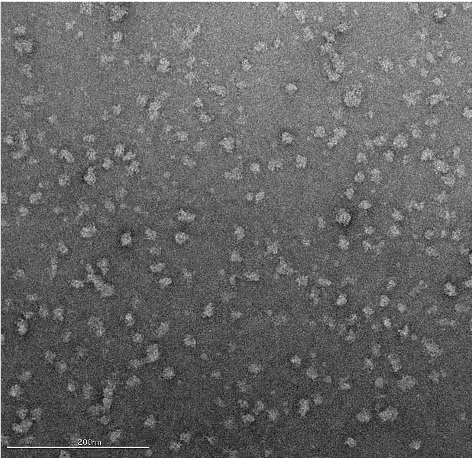
Negative-stain transmission electron microscopy can provide an early view of particle quality before a single-particle cryo-EM study. The method offers high image contrast and can help assess whether a sample appears monodisperse, aggregated, dissociated, heterogeneous, or present at an appropriate concentration. For some complexes, however, the dilution used during negative-stain preparation can shift the binding equilibrium and make the sample less representative of the material intended for vitrification.
The practical question is therefore conditional: will negative stain reduce uncertainty about the sample, or could preparation alter the complex enough to obscure the relevant state? The answer depends on sample behavior, binding affinity, dissociation kinetics, stoichiometry, concentration, and the decision the imaging study must support.
Negative Stain Electron Microscopy
Negative-stain EM uses an electron-dense stain to surround and lightly coat biological particles on a support film. The stain increases contrast in the transmission electron microscope and can make particle size, approximate shape, distribution, and sample heterogeneity easier to inspect. Because the specimen is dried and embedded in stain, the resulting images should be interpreted as a screening or structural-characterization view rather than a direct representation of particles in vitreous ice [1, 2].
What negative stain can reveal
- Whether particles appear uniform and monodisperse.
- Whether particle size and approximate shape are consistent with expectations.
- Whether the sample appears too sparse, crowded, aggregated, or partly dissociated.
- Whether multiple particle populations or conformational states may be present.
- Whether the sample warrants deeper image analysis or a change in preparation conditions before vitrification.
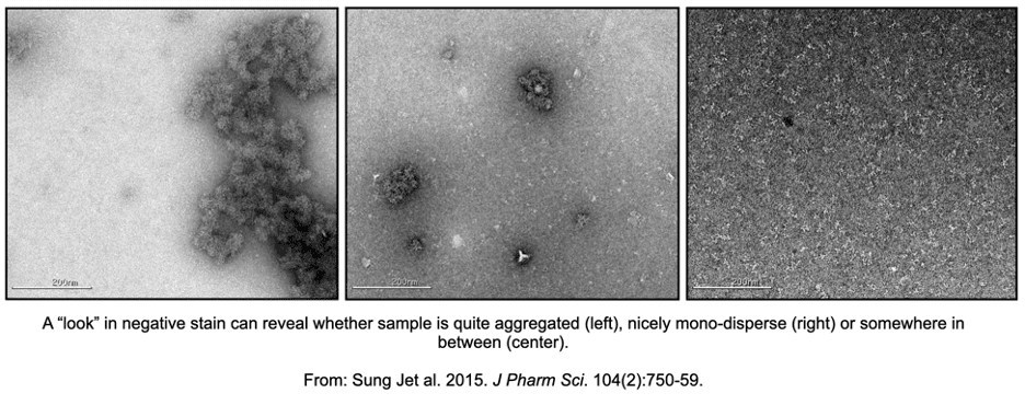
What Negative Stain Reveals During Initial Sample Assessment
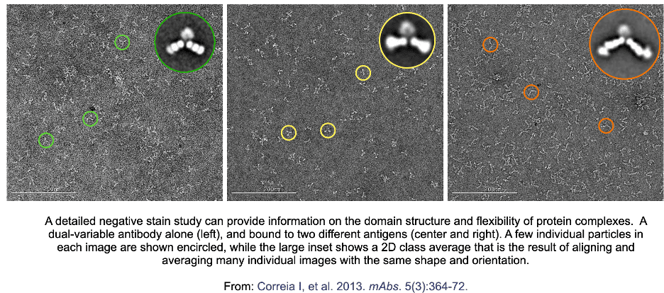
A small set of micrographs can support an initial assessment of particle distribution and sample integrity. A more detailed study can examine domain organization, antibody flexibility, or whether a protein complex forms as expected. The stain thickness and particle distribution should be optimized so particles remain visible without excessive overlap or overly sparse fields.
How 2D classification extends the assessment
For deeper analysis, individual particle images can be selected, aligned, and grouped by similar shape and orientation through 2D classification. Averaging many particles within a class improves the signal relative to noise and can clarify domain organization, flexibility, compositional variation, or binding-state differences [1, 4]. The result remains dependent on preparation quality and the particle views represented in the data.
Typical Protein Concentration for Cryo-EM Grids and Negative Stain
Concentration targets are practical starting points and should be adjusted for the particle, buffer, grid, ice behavior, and analytical goal. In the workflow described here, negative-stain samples are commonly diluted to approximately 10 to 50 micrograms per milliliter before grid preparation. Cryo-EM grids are discussed at approximately 0.5 to 4 milligrams per milliliter. These ranges place negative-stain preparation roughly one to two orders of magnitude below the cryo-EM concentration range in the worked example.
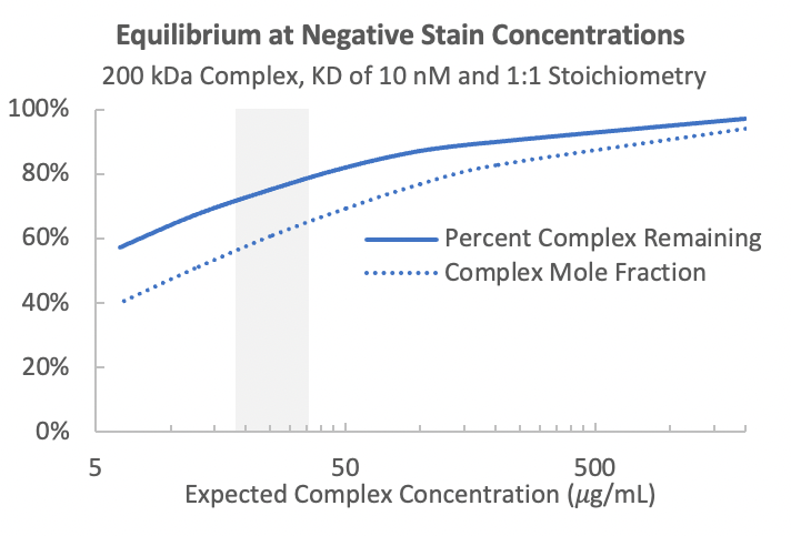
That difference can be useful when sample is scarce, but lower concentration can also perturb a reversible complex. A concentration that produces clear negative-stain particles may still change the fraction of assembled complex relative to the higher-concentration material used for vitrification.
How Dilution Can Affect Protein-Complex Stability
For a simple reversible 1:1 interaction between components A and B, the dissociation constant can be written as Kd = [A][B] / [AB]. When total concentration falls, the equilibrium can shift toward the unbound components even when the interaction is considered relatively high affinity.
An illustrative calculation for a 200 kDa complex with a dissociation constant of 10 nM and 1:1 stoichiometry estimates that, at approximately 30 micrograms per milliliter, only 75 percent of the complex remains intact. The complex mole fraction is 61 percent because free A, free B, and complex AB all contribute to the particle population. This is a worked example rather than a universal prediction. Larger complexes, lower-affinity interactions, more complex stoichiometries, and multiple dissociation constants can produce different behavior.
Negative Stain EM Resolution and Limitations
Negative-stain EM is generally used for lower-resolution particle assessment and structural characterization. A peer-reviewed methods paper describes a practical resolution limit around 18 to 20 angstroms, although the interpretable detail depends on the specimen, staining method, support film, particle orientation, and image processing [2]. This range should be treated as a guideline rather than a guaranteed outcome.
- Drying, adsorption to carbon, and stain depth can alter particle appearance.
- Preferred orientation on the support film can limit the views available for reconstruction.
- High contrast can reveal smaller particles, while staining artifacts can complicate interpretation.
- A class average can improve visibility of recurring features, but it cannot recover states that are absent or destabilized during preparation.
When to Use Negative Stain Before Cryo-EM
Negative stain is often informative when the main uncertainty concerns aggregation, particle integrity, approximate size and shape, heterogeneity, flexibility, complex formation, or whether a sample is ready for more resource-intensive vitrified screening [5, 6]. It is especially useful when the preparation is unlikely to alter the state that matters for the study.
- Use an initial negative-stain assessment when a rapid visual check can change the sample-preparation plan.
- Extend to 2D classification when domain organization, conformational variation, or binding-state differences are relevant.
- Compare the intended negative-stain concentration with affinity and stoichiometry data before assuming the complex will remain assembled.
- Document stain behavior, particle distribution, preferred orientations, and visible artifacts when interpreting the result.
When to Skip Negative Stain and Go Directly to Cryo-EM
Direct vitrification may be more appropriate when the complex is expected to dissociate at negative-stain concentrations, when dilution is difficult to model, or when the relevant state must be evaluated close to the concentration used for cryo-EM grid preparation. Screening a vitrified sample can require more time and sample than negative stain, so the decision should be tied to the risk of obtaining misleading negative-stain data.
For protein and small-molecule complexes, using a 5- to 10-fold molar excess of the small molecule can shift the equilibrium toward complex formation when scientifically appropriate. For protein-protein complexes, options include calculating the expected dilution effect or preparing the grid immediately after dilution when the complex has a slow dissociation rate. These approaches are sample-specific and should be evaluated against the study objective.
A Practical Decision Framework
- Define the decision: confirm particle quality, evaluate complex formation, characterize heterogeneity, or proceed toward structure determination.
- Review sample constraints: available quantity, concentration, buffer, stability, affinity, stoichiometry, and dissociation kinetics.
- Estimate whether negative-stain dilution could change the assembled fraction or particle population.
- Choose the least disruptive method that can answer the immediate question.
- Set an explicit checkpoint for moving from negative stain to vitrified screening or for proceeding directly to cryo-EM.
Frequently Asked Questions
What is a negative stain?
A negative stain is an electron-dense material applied around biological particles on a support film. It increases contrast, so particle outlines, distribution, approximate shape, aggregation, and heterogeneity can be assessed by TEM.
What is negative stain electron microscopy?
Negative-stain electron microscopy is a TEM method in which particles are absorbed to a grid, surrounded by stain, blotted, and dried. It is commonly used for sample assessment and lower-resolution structural characterization.
What is the typical protein concentration for cryo-EM?
In the workflow described here, a practical cryo-EM grid preparation range is approximately 0.5 to 4 milligrams per milliliter. The appropriate concentration remains sample- and grid-dependent.
What is the typical protein concentration for cryo-EM grids compared with negative stain?
Negative-stain preparation is described at approximately 10 to 50 micrograms per milliliter, while cryo-EM grid preparation is described at approximately 0.5 to 4 milligrams per milliliter. The lower negative-stain concentration can affect reversible complexes.
Can negative stain be used before cryo-EM?
Yes. Negative stain can support initial assessment of aggregation, particle integrity, approximate size and shape, heterogeneity, and complex formation before vitrification. It is most informative when preparation is unlikely to destabilize the state of interest.
When should you skip negative stain?
Direct cryo-EM screening may be appropriate when dilution is expected to dissociate the complex, when the dilution effect is difficult to predict, or when the relevant state must be assessed near the concentration intended for vitrification.
What resolution can negative stain EM achieve?
A peer-reviewed methods paper describes a practical limit around 18 to 20 angstroms. Treat that range as a guideline because specimen behavior, stain, support film, particle orientation, and processing affect interpretable detail.
How does 2D classification help negative stain EM?
2D classification groups aligned particle images with similar shapes and orientations. Class averaging improves signal relative to noise and can clarify recurring domain arrangements, flexibility, heterogeneity, or binding states.
Discuss a Negative-Stain and Cryo-EM Strategy
TrueCourse can help evaluate whether negative-stain TEM, direct vitrified screening, or a staged combination is appropriate for a protein or protein complex. Planning should account for the scientific question, available sample, target concentration, binding equilibrium, dissociation kinetics, stoichiometry, particle behavior, and the decision the imaging data must support.
[ CTA button ]
Peer-reviewed references cited in the draft
[1] Ohi, M., Li, Y., Cheng, Y., & Walz, T. (2004). Negative staining and image classification: powerful tools in modern electron microscopy. Biological Procedures Online, 6, 23-34. https://doi.org/10.1251/bpo70
[2] Scarff, C. A., Fuller, M. J. G., Thompson, R. F., & Iadaza, M. G. (2018). Variations on negative stain electron microscopy methods: tools for tackling challenging systems. Journal of Visualized Experiments, 132, e57199. https://doi.org/10.3791/57199
[3] Sung, J. J., Pardeshi, N. N., Mulder, A. M., et al. (2015). Transmission electron microscopy as an orthogonal method to characterize protein aggregates. Journal of Pharmaceutical Sciences, 104, 750-759. https://doi.org/10.1002/jps.24157
[4] Correia, I., Sung, J., Burton, R., et al. (2013). The structure of dual-variable-domain immunoglobulin molecules alone and bound to antigen. mAbs, 5, 364-372. https://doi.org/10.4161/mabs.24258
[5] Cheng, Y., Grigorieff, N., Penczek, P. A., & Walz, T. (2015). A primer to single-particle cryo-electron microscopy. Cell, 161, 438-449. https://doi.org/10.1016/j.cell.2015.03.050
[6] Cianfrocco, M. A., & Kellogg, E. H. (2020). What could go wrong? A practical guide to single-particle cryo-EM: from biochemistry to atomic models. Journal of Chemical Information and Modeling, 60, 2458-2469. https://doi.org/10.1021/acs.jcim.9b01178
.png)
.png)
.png)