Seeing Through the Fog: Using Cryo-TEM to Characterize LNPs from Formulation Through Manufacturing
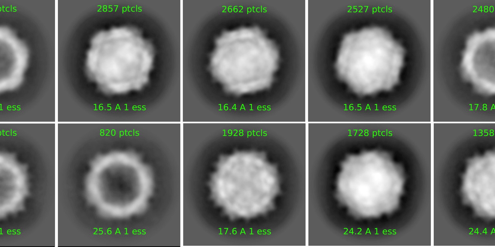
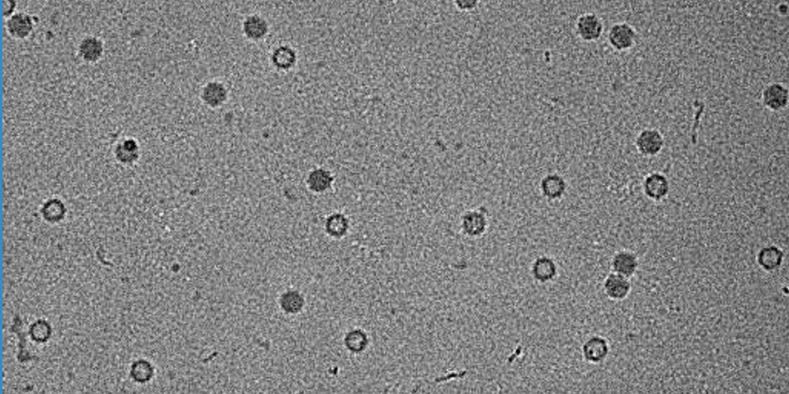
Bulk assays provide essential measurements for lipid nanoparticle development, but an average size or encapsulation value cannot describe every particle in a formulation. Cryo-transmission electron microscopy adds direct, particle-level insight into LNP size distributions, payload organization, morphology, lamellarity, bleb formation, aggregation, and free nucleic acid.
Following LNP Structure Through Development
This white paper follows an mRNA LNP formulation across mixing platforms, deliberate formulation and manufacturing changes, an unexpected process deviation, and refrigerated storage. Quantitative cryo-TEM image analysis is compared with DLS and RiboGreen results to show where batches that appear similar by bulk measurement can differ structurally.
What You’ll Learn in the White Paper
- Which LNP attributes cryo-TEM measures on a particle-by-particle basis
- How mixing platform, buffer, purification method, and payload can influence morphology
- How direct imaging can reveal process deviations, free mRNA, and storage-related changes
- How sampling and consistent classification support meaningful batch comparisons
Download the white paper to explore how cryo-TEM can inform LNP formulation screening, process development, scale-up, comparability, and stability assessment.